本文已于2026年3月在线发表于Journal of Nuclear Medicine and Molecular Imaging杂志。
文章链接(Article Link):https://mednexus.org/doi/epdf/10.65457/JNMMI-2026-00xx-6
引用本文:Meng X, Wang R. 18F-FDG PET/CT in a case of primary pulmonary epithelioid angiosarcoma. J Nucl Med Mol Imaging 2026
How to cite this article: Meng X, Wang R. 18F-FDG PET/CT in a case of primary pulmonary epithelioid angiosarcoma. J Nucl Med Mol Imaging 2026
摘要:原发性肺上皮样血管肉瘤(PPA)是一种起源于血管内皮的恶性血管源性肿瘤,极为罕见,临床症状和影像学特征缺乏特异性,极易被误诊,目前病理诊断是唯一金标准。其发病机制尚未完全阐明,据有关报道,可疑发病危险因素是放疗、二氧化钍、聚氯乙烯、氡、开胸剂、铜矿粉尘、乳房切除术、慢性脓胸和结核病等。本例患者为女性,无相关危险因素接触史,属极罕见病例,起初被误诊为肺部感染。
Primary pulmonary epithelioid angiosarcoma (PPA) is a rare malignant tumor originating from vascular endothelium. It lacks specific clinical manifestations and imaging features, making it prone to misdiagnosis. Pathological biopsy combined with immunohistochemistry is the only gold standard for confirmation.
病历资料:患者女,54岁,主因“间断性咳嗽、咯血、伴有黑便1月余”入院。查体贫血貌。实验室检查:CEA 7.99μg/L,CA125 153.50U/mL,NSE 31.26ng/mL,CYFRA21-1 5.07ng/mL,血清铁蛋白 541.70ng/mL;中性粒细胞0.920,白细胞计数15.21×109/L,红细胞计数1.95×1012/L,血红蛋白测定58g/L,白细胞介素-6 75.14pg/mL,C-反应蛋白8.425mg/dL;凝血功能各项指标异常。PET/CT:双肺多发片状及结节状实变影,周围伴大片状磨玻璃影,代谢不均匀增高;脾脏增大伴弥漫性代谢增高;扫描范围内骨髓弥漫性代谢增高。病理穿刺:低分化上皮样恶性肿瘤伴大片坏死;免疫组化:CD31(+),ERG(+),FLI-1(+),CK(肺泡上皮+,肿瘤细胞-),CK5(-),p63(-),p40(-),TTF-1(-),NapsinA(-),ALK(Ventana)(-),Brg-1(+),PDL1(SP263)(TPS:50%);病理诊断为:肺上皮样血管肉瘤。
This case involves a 54-year-old female patient with no history of exposure to relevant risk factors (an extremely rare scenario) who was admitted due to "intermittent cough, hemoptysis, and melena for more than 1 month." Physical examination revealed anemic appearance. Laboratory tests indicated elevated tumor markers, severe anemia, and abnormal coagulation function. PET/CT showed multiple patchy and nodular consolidation shadows in both lungs with ground-glass opacities and increased metabolism, accompanied by splenomegaly with diffusely increased metabolism and diffusely increased metabolic activity in the visualized bone marrow. The diagnosis was confirmed by pathological puncture and immunohistochemistry (CD31 (+), ERG (+), FLI-1 (+), PD-L1 expression rate of 50%). Given the patient's poor general condition and inability to tolerate chemotherapy, single-agent immunotherapy was ultimately recommended.
讨论:PPA极为罕见,临床症状无特异性,可见咳嗽、咳血等,发病年龄多在40岁及以上,中老年男性多见,复发和转移率高,预后较差。最终确诊需依靠病理组织学检查及免疫组织化学分析,CD31、EGR、FLI-1是近年来研究发现的高度敏感和特异的免疫组化标志物,该例患者此三项标记物均为阳性表达。PPA治疗选择目前主要包括手术、放疗和化疗。此外,有研究表明靶向药物和免疫治疗对血管肉瘤有效。但由于其侵袭性强,确诊时多已晚期,疗效欠佳。本病例一般状况差、有咯血症状,考虑到化疗不能耐受及靶向治疗的副作用,结合免疫组化PDL1(SP263)(TPS:50%),联合会诊后建议给予单药免疫治疗。既往发表的个案中极少有关于PPA FDG PET/CT成像的相关报道,目前看来其对于鉴别原发和转移提供了价值,但对于鉴别PPA与其他恶性肿瘤仍缺乏特异性。
This case report supplements the clinical data of PPA in females without risk factors, enriching the case spectrum of this rare disease. It confirms the key value of pathological biopsy combined with CD31, ERG, and FLI-1 immunohistochemical markers for the diagnosis of PPA. It provides practical reference for immunotherapy in advanced PPA patients with high PD-L1 expression. Additionally, it clarifies the value of PET/CT in distinguishing primary from metastatic lesions and assessing the extent of PPA, while pointing out its limitations in differentiating PPA from other malignant tumors or infectious lesions, thus offering insights for clinical imaging diagnosis.
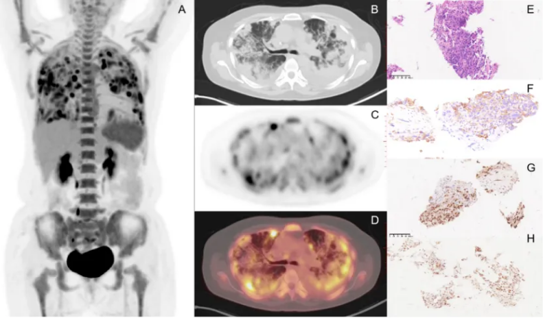
Figure 1. (A) The maximal intensity projection(MIP); (B-D) The 18F-FDG PET/CT shows: Bilateral multiple patchy and nodular consolidation shadows, surrounded by large patchy ground-glass opacities, with unevenly increased metabolism, maximum SUVmax on PET: 11.5; (E) Specimen stained with hematoxylin and eosin showed histological findings of epithelioid angiosarcoma; (F) IHC: Tumor cells showing immunopositivity to CD31; (G) Tumor cells showing immunopositivity to EGR; (H) Tumor cells showing immunopositivity to FLI-1.
作者简介